



Microscope Breakthrough Unveils Real-Time Flu Virus Invasion, Offering New Insights Into Disease Prevention and Control
 By
Noah Bennett
By
Noah Bennett
| IN A NUTSHELL |
|
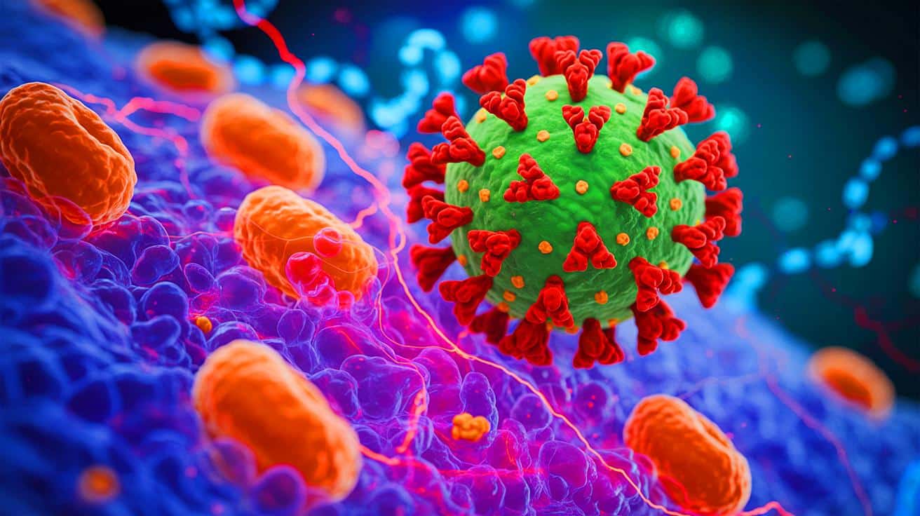
Every winter, as temperatures drop, the flu virus reemerges as a familiar and unwelcome visitor. This virus, carried by tiny droplets in the air, exploits our respiratory system, targeting cells essential for our survival. Its attack is facilitated by two molecular keys, hemagglutinin (HA) and neuraminidase (NA), which enable it to infiltrate cells. The invasion process, known as endocytosis, has long been a challenge to observe due to its rapid and minuscule nature. However, a revolutionary new imaging technique has granted scientists unprecedented insight into this microscopic drama, potentially reshaping our understanding of viral infections and paving the way for innovative treatments.
The Dance of Virus and Cell
The intricate interaction between the flu virus and human cells has been likened to a dance, a metaphor used by Yohei Yamauchi at ETH Zurich. This analogy captures the dynamic relationship where the virus and cell engage in a complex battle. The virus, equipped with HA and NA proteins, attaches to sialic acids on the cell surface. It then glides along until the cell engulfs it.
What has remained elusive, until now, is the role of the cell in this encounter. Traditionally viewed as a passive victim, the cell is now seen as an active participant. The cell’s membrane stretches and shifts, seemingly attempting to control the viral invasion. This new perspective challenges previous assumptions and underscores the complexity of viral infections.
The observation of these interactions was made possible by combining atomic force microscopy (AFM) and fluorescence microscopy, creating ViViD-AFM. This innovative tool allows scientists to view the nanoscale processes in real time, offering a clearer picture of the viral invasion and the cell’s response.
A New Lens on Viral Invasion
The development of ViViD-AFM represents a significant leap forward in microscopy. It enables researchers to observe the flu virus without traditional fluorescent tags, which can alter the behavior of the virus. By capturing both the physical shape of the cell and the fluorescent signals from the virus, this method provides a detailed view of the interaction between viral proteins and the cell surface.
One of the critical discoveries using this technique is the role of actin, a protein that shapes the cell. The virus requires larger protrusions on the cell surface, formed by actin, to successfully enter. These findings suggest that the mechanism of viral entry is akin to other cellular processes, further deepening our understanding of cell biology.
The ability to study these interactions in living cells offers immediate feedback on how antiviral drugs perform, potentially accelerating the development of effective treatments. ViViD-AFM could also be applied to other viruses and aid in the study of vaccine-cell interactions.
Implications for Drug Development
The potential applications of ViViD-AFM extend far beyond influenza research. By providing a “window” into the cellular world, this tool offers invaluable insights into how drugs interact with cells. Researchers can observe, in real time, how medicines influence viral behavior and cellular responses.
This capability could revolutionize drug development, allowing for more targeted and effective treatments. By understanding precisely how drugs affect cellular processes, scientists can tailor therapies that are more efficient and have fewer side effects.
Furthermore, this technique could facilitate the study of nanoparticles used in drug delivery. It may uncover how these particles penetrate cells, offering clues to optimize their design for better therapeutic outcomes.
A Versatile Tool for Future Research
ViViD-AFM is poised to become an essential tool in biological research. Its versatility allows scientists to investigate a wide range of cellular interactions, from virus attachment to the release of extracellular vesicles. The detailed insights it provides could lead to groundbreaking discoveries in virology, medicine, and cellular biology.
As researchers continue to explore its capabilities, ViViD-AFM may unlock new pathways in understanding cell-virus interactions. This could lead to innovations in vaccine development, antiviral therapies, and even cancer research, where cell behavior is crucial.
The findings of this study, published in the journal PNAS, mark a significant milestone in microscopy and virology. The insights gained from this research hold the potential to transform our approach to combating viral infections and enhance our overall understanding of cellular processes.
As the scientific community delves deeper into the applications of ViViD-AFM, one question remains: How will these advancements in microscopy shape the future of medicine and our ability to combat viral threats?
Continue reading